
smile-design
How to Build a Full Smile Makeover Plan That Works
Full smile makeover plan explained: how to properly sequence Invisalign, whitening, and veneers together for the best result.
Sep 22, 20266 min read



CBCT imaging for implant planning explained simply: it's a 3D X-ray that captures your jawbone in far more detail than a standard dental X-ray, giving your full mouth reconstruction team a complete map before any implant is placed. That map is what makes precise, predictable implant planning possible.
Dental implants have a success rate of 95 to 98% over 10 years, according to the Journal of Oral and Maxillofacial Surgery, and accurate planning is a major reason why. This guide explains what CBCT imaging actually shows, why it matters for implant cases specifically, and what to expect if it's part of your treatment plan for full-arch or single-implant placement.
Patients researching implants for the first time often assume the planning happens mostly during the surgery itself, with the dentist making judgment calls in the moment based on what they see and feel. In reality, the bulk of the critical planning decisions happen well before that, often days or weeks ahead of any procedure, based on the detailed 3D picture CBCT provides. Understanding that shift in when decisions actually get made helps explain why imaging technology plays such a central role in modern implant treatment.
CBCT stands for cone beam computed tomography. It's a type of 3D X-ray that scans your jaw from multiple angles and combines the images into a detailed three-dimensional model. It shows structures a standard 2D X-ray simply can't capture.
Unlike a traditional dental X-ray, which produces a flat, two-dimensional image, CBCT works differently. It rotates a cone-shaped beam around your head to build a full 3D reconstruction of your jawbone, teeth, nerves, and sinus structures. The scan itself takes less than a minute. The equipment then assembles hundreds of individual images into the final 3D model afterward.
| Feature | Standard 2D X-Ray | CBCT 3D Imaging |
|---|---|---|
| Image type | Flat, two-dimensional | Full three-dimensional model |
| Bone width visible | No | Yes |
| Nerve and sinus position | Approximate | Precise |
| Surgical guide creation | Not possible | Possible |
Radiation exposure from CBCT is low. It's generally comparable to or only somewhat higher than a full set of traditional dental X-rays, and well below the exposure of a medical CT scan. That makes it a practical tool for routine implant planning rather than something reserved for rare cases.
The technology has become common enough in dental offices that many patients considering implants encounter it as a routine part of the planning process rather than something unusual. Most practices offering implant treatment either have CBCT equipment on-site or coordinate with an imaging center to obtain the scan before finalizing a treatment plan, a workflow covered further in the ADA's patient resources on dental implants.
CBCT imaging is used for implant planning because it shows the exact height, width, and density of your jawbone in 3D. It also pinpoints the precise location of nerves and sinuses that must be avoided during placement. A flat X-ray can't provide that depth of detail.
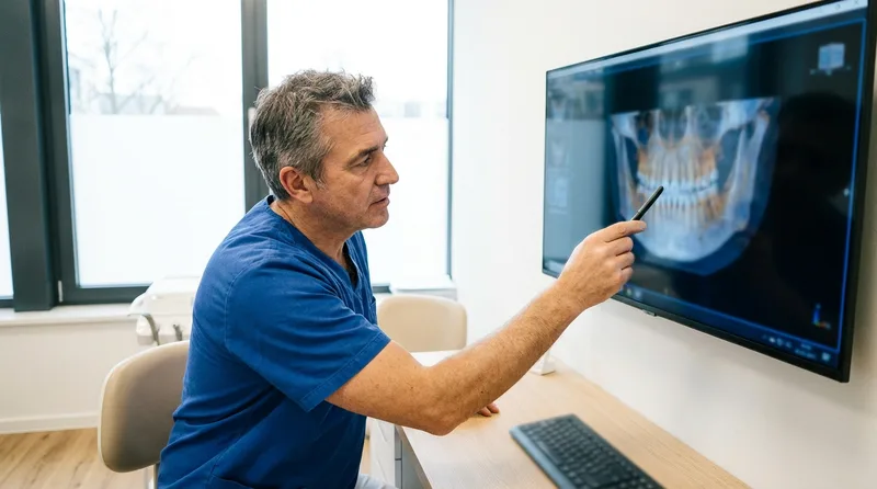
Placing a dental implant successfully depends on having enough bone in the right location. It also depends on knowing exactly where sensitive structures like the inferior alveolar nerve or sinus floor sit relative to that bone. A 2D X-ray can suggest general bone height but can't reliably show width or the true 3D position of nearby anatomy. CBCT removes much of that guesswork, which is part of why it's become a standard part of implant planning rather than an optional add-on.
This matters most in the lower jaw near the nerve canal and in the upper jaw near the sinus floor, where a small miscalculation in depth or angle can have real consequences. Having a precise 3D map before treatment turns what used to require more conservative estimating into a plan built on actual measurements specific to your anatomy.
CBCT imaging shows bone width, bone density, the exact 3D position of nerves and sinuses, and the precise angle and depth available for implant placement, none of which a standard 2D X-ray can capture with the same accuracy. This extra dimension changes planning decisions.
A regular X-ray might show that bone appears present at a proposed implant site, but it can't confirm whether that bone is wide enough to support the implant safely, since a 2D image flattens depth into a single plane. CBCT reveals that missing dimension, which is often the difference between a straightforward placement and one that needs bone grafting first to build up insufficient width or height.
Density matters just as much as raw dimensions. Bone that looks adequate in height and width can still be too soft or porous to support an implant reliably, and CBCT's density readings help identify that before surgery rather than discovering it mid-procedure. That advance knowledge lets your dental team plan for grafting, choose a different implant size, or adjust positioning ahead of time instead of improvising in the moment.
CBCT imaging allows your dentist or oral surgeon to plan the exact position, angle, and depth of each implant digitally before surgery, sometimes using that data to create a surgical guide that helps place implants precisely as planned. This reduces surprises during the actual procedure.
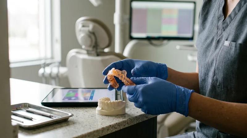
With a detailed 3D model in hand, cases that might otherwise have required an in-surgery decision, like adjusting an implant's angle after encountering unexpected bone density, can often be resolved during planning instead. Surgical guides built from CBCT data essentially create a template that keeps each implant tracking to the pre-planned position, angle, and depth throughout placement.
This level of pre-surgical planning also shapes conversations about the overall treatment timeline. If the scan reveals insufficient bone in a specific area, that's identified and addressed, often with grafting, well before the implant surgery itself rather than as an unexpected delay discovered mid-procedure. Patients tend to appreciate knowing the full scope of their treatment plan upfront rather than learning about additional steps partway through.
Considering dental implants?
A CBCT-guided consultation gives you a clear picture of your bone structure and candidacy before any decisions are made.
Schedule a Consultation →A CBCT scan takes less than a minute and involves standing or sitting still while the machine rotates around your head, similar in feel to a panoramic dental X-ray. There's no discomfort involved and no need for sedation.
You'll be asked to remove metal jewelry, glasses, or anything else that could interfere with the image, and to bite on a small positioning piece to keep your jaw steady during the scan. Once complete, the images are processed into a 3D model that Dr. Curley or the coordinating specialist reviews with you, often on the same visit, to walk through what the scan shows for your specific case.
There's no anesthetic and nothing invasive about the scan itself. Most patients describe it as quick and unremarkable, closer to a routine imaging step than anything resembling treatment. Reviewing the resulting 3D model together afterward tends to be the more memorable part of the visit, since it's often the first time patients see their own jawbone structure this clearly.
Most implant cases benefit from CBCT imaging, though the exact need depends on the number of implants planned, whether bone grafting might be involved, and the complexity of the case. Single, straightforward implant cases sometimes rely on more limited imaging.
Full-arch cases, cases near the sinus or major nerves, and any situation where bone volume is uncertain almost always call for CBCT imaging given how much planning detail it provides. For patients considering our implant options compared to other tooth-replacement approaches, CBCT often factors into that broader planning conversation as well, since it clarifies exactly what your jawbone can support before a final recommendation is made.
The decision ultimately rests with your dentist or the coordinating specialist based on your specific anatomy and treatment goals. A straightforward single-tooth case with clearly adequate bone on a standard X-ray might proceed with more limited imaging, while anything involving multiple implants, a history of bone loss, or proximity to sensitive structures generally warrants the fuller picture CBCT provides.
CBCT imaging improves the accuracy of implant placement by giving the planning team a complete picture of bone and anatomy beforehand, which reduces the risk of complications tied to inadequate bone or unexpected proximity to nerves. Precise planning supports the strong long-term outcomes implants are known for.
Dental implants have a success rate of 95 to 98% over 10 years, according to the Journal of Oral and Maxillofacial Surgery, and thorough imaging-based planning is one of the contributing factors behind that consistency. While no imaging technology eliminates every variable in implant treatment, reducing the guesswork around bone structure and anatomy meaningfully lowers the chance of avoidable complications.
It's worth putting this in perspective: implant treatment has always had a strong track record, even before CBCT became standard. What the technology adds is more precision and fewer surprises along the way, particularly for complex cases involving multiple implants or challenging anatomy, where the margin for error matters most. For straightforward, single-tooth cases with generous bone volume, the value is still real but somewhat less dramatic than in a full-arch or bone-graft scenario.
Patients sometimes ask whether skipping CBCT to save time or cost is a reasonable option. For most implant cases, the answer is no, since the risk of an avoidable complication or an unexpectedly difficult surgery generally outweighs whatever is saved by skipping detailed imaging. The scan itself is quick and low-radiation, so the trade-off rarely favors skipping it.
CBCT imaging for implant planning explained comes down to a simple idea: seeing more before treatment begins means fewer surprises during it. It's a routine, low-radiation step, generally in line with general dental imaging safety guidance from the CDC, that gives both patient and dental team a clear, shared picture of what's possible. Results may vary. Please consult with your dentist at Susan J. Curley DDS for personalized treatment recommendations.
If you've been considering dental implants and want to understand how your case would actually be planned, a consultation that includes CBCT imaging is the clearest way to find out.
Considering Dental Implants?
Schedule a consultation with Dr. Curley in Wall Township and see exactly what CBCT imaging shows for your case.
Book Appointment →Have questions before booking?
Contact Our Wall Township Office →Written By
Dentist
Full smile makeover plan explained: how to properly sequence Invisalign, whitening, and veneers together for the best result.
Digital smile design mockup explained honestly for patients: how it is created, why it is trustworthy, and what it shows you.
Smile design for a gummy smile: what actually causes it and which treatment options can genuinely help you feel confident.
Book your visit with Dr. Curley online or call us, and we'll take it from there.